Article Text

Abstract
Objectives To conduct a systematic review of changes in lung function in relation to presence of pleural plaques in asbestos-exposed populations.
Methods Database searches of PubMed and Web of Science were supplemented by review of papers’ reference lists and journals’ tables of contents. Methodological features (eg, consideration of potential confounding by smoking) of identified articles were reviewed by ≥two reviewers. Meta-analyses of 20 studies estimated a summary effect of the decrements in per cent predicted (%pred) forced vital capacity (FVC) and forced expiratory volume in 1 s (FEV1) associated with presence of pleural plaques.
Results Among asbestos-exposed workers, the presence of pleural plaques was associated with statistically significant decrements in FVC (4.09%pred, 95% CI 2.31 to 5.86) and FEV1 (1.99%pred, 95% CI 0.22 to 3.77). Effects of similar magnitude were seen when stratifying by imaging type (X-ray or high-resolution CT) and when excluding studies with potential methodological limitations. Undetected asbestosis was considered as an unlikely explanation of the observed decrements. Several studies provided evidence of an association between size of pleural plaques and degree of pulmonary decrease, and presence of pleural plaques and increased rate or degree of pulmonary impairment.
Conclusions The presence of pleural plaques is associated with a small, but statistically significant mean difference in FVC and FEV1 in comparison to asbestos-exposed individuals without plaques or other abnormalities. From a public health perspective, small group mean decrements in lung function coupled with an increased rate of decline in lung function of the exposed population may be consequential.
- pleural plaque
- FVC
- FEV1
This is an Open Access article distributed in accordance with the Creative Commons Attribution Non Commercial (CC BY-NC 4.0) license, which permits others to distribute, remix, adapt, build upon this work non-commercially, and license their derivative works on different terms, provided the original work is properly cited and the use is non-commercial. See: http://creativecommons.org/licenses/by-nc/4.0/
Statistics from Altmetric.com
Introduction
Asbestos is the generic name for a group of naturally occurring silicate minerals that crystallise in long thin fibres. Asbestos has been used in a wide range of applications such as insulation, friction materials and textiles; worldwide asbestos usage peaked around the 1970s and has since declined due to regulations enacted to decrease or prevent exposure.1 However, such regulations vary by region and country, and considerable amounts of asbestos are still used today—for example, the US Geological Survey estimated that the worldwide production of asbestos was nearly 2 million metric tons in 2012, and that the USA consumed 1020 metric tons of asbestos for applications (almost exclusively in the chloralkali industry and roofing products).2 Further, naturally occurring asbestos is wide spread in the USA.3 Asbestos exposure and subsequent health effects continue to be a public health concern.
Asbestos has long been known to cause mesothelioma, along with lung and various other cancers (eg, laryngeal and ovarian).1 Asbestos is also known to cause various non-cancer effects in the lung (eg, asbestosis) and/or the pleura (eg, pleural plaques, diffuse pleural thickening (DPT)). Pleural plaques are one of the earliest and most common manifestations of asbestos-related disease. Pleural plaques are lesions in the tissue surrounding the lungs and lining the chest cavity.4
Pleural plaque prevalence increases with increasing time since first exposure; in some cohorts, after decades of follow-up, the prevalence of pleural plaque is over 80%.5 ,6 The impact of pleural plaques has been debated in the literature. The American Thoracic Society (ATS),4 stated that “Although pleural plaques have long been considered inconsequential markers of asbestos exposure, studies of large cohorts have shown a significant reduction in pulmonary function attributable to the plaques, averaging about 5% of FVC, even when interstitial fibrosis (asbestosis) is absent radiographically…Decrements, when they occur, are probably related to early subclinical fibrosis.” The American College of Chest Physicians (ACCP)7 published a Delphi study conducted to gauge consensus among published asbestos researchers, and found that these researchers rejected the statement that “Pleural plaques alter pulmonary function to a clinically significant degree.” However, neither the ATS nor the ACCP statements were based on a formal systematic review of the literature. Recently, Wilken et al8 performed a systematic review and meta-analysis, examining pulmonary function in relation to the combined category of pleural plaques and/or DPT. DPT is thought to be a more severe health outcome compared with pleural plaques, and associated with more severe decrements in lung function.4 Mixing the two end points does not allow evaluation of the effect of pleural plaques alone.
Our objective was to conduct a systematic evaluation of cross-sectional and longitudinal studies examining the relationship between pleural plaques and lung function, focusing on changes in per cent predicted (%pred) forced vital capacity (FVC) and forced expiratory volume in 1 s (FEV1), the most commonly reported measures in the identified studies. We considered X-ray studies and newer high-resolution CT (HRCT) studies.
Methods
Literature search strategy
The search was conducted on 25 September 2013 using the PubMed and Web of Science databases with no publication date limitations; the search strategy (including search strings) is summarised in online supplementary figure 1, with additional details of the process described below.
Standardised guidelines for defining plaques using radiographic evaluation are provided by the International Labour Organization (ILO), and have changed over time. The 1980 ILO guidelines9 defined circumscribed pleural thickening (ie, pleural plaques). The 2000 ILO revision10 defined a new category of localised pleural thickening (LPT) comprising only those plaques with width of at least 3 mm (intended to reduce the number of false-positive findings), and including plaques found on sites other than the chest wall (eg, diaphragm). Both pleural plaques as defined by the earlier ILO guidelines, and LPT as defined by the 2000 guidelines, were included in our literature search. There are no standardised guidelines similar to the ILO for defining pleural plaques using HRCT; thus, we used the authors’ descriptions of the definition for pleural plaques.
The searches yielded 184 hits in PubMed, and 183 hits in Web of Science; after excluding duplicate citations, 262 remained for further review. On the basis of a title and abstract screen, 105 citations were excluded because they were not directly relevant to the study question (eg, no pulmonary function measurements). The remaining 157 citations were selected for full-text review by a group of three reviewers to determine if they contained data addressing our study question. Each paper was reviewed independently by two of the three reviewers. In cases of disagreements or uncertainty (eg, questions about the definition of pleural abnormality used), the third reviewer also reviewed the paper and participated in the consensus building discussions. Studies were also excluded at this step if the analysis group included individuals with DPT or was based on undefined pleural abnormalities (n=21), or if they included individuals with parenchymal abnormalities (defined as X-ray profusion score >1/0, or HRCT evidence of parenchymal abnormality) without presenting a stratified analysis showing the results for the effect of pleural plaques in the absence of parenchymal abnormality (n=7). Thirty studies were selected for inclusion through this process, and eight additional references were identified through (1) a review of references in reviews and in the identified primary source studies and (2) by searching the Table of Contents of relevant journals for newly released papers (September–December 2013) of selected journals (see online supplementary material) for a total of 38 primary source studies. All of the X-ray studies used in these meta-analyses stated that they used the outcome of pleural plaques as defined by the 1980 ILO guidelines; no studies reported LPT as defined by the 2000 ILO guidelines. If more than one publication presented data on the same study participants or on a subset of the study participants, or provided additional methodological details about a study, these publications are treated as related (with one entry in the summary tables and analysis). Some studies presented both longitudinal and cross-sectional data from the same study population; the longitudinal and cross-sectional results were considered separately.
In the next step of this review process, each of the selected studies was evaluated for attributes related to study methods. Again, two of the three reviewers independently abstracted information pertaining to: selection of participants, protocols for X-ray or HRCT readings, protocols for spirometry measurements, analytic approach and consideration of smoking as a potential confounder (see online supplementary table S1). These criteria were defined a priori. This information was not used as a basis for exclusion, but rather to identify studies with limitation(s) of sufficient magnitude to potentially affect the interpretation of the study results.
For the purpose of developing a summary effect estimate across studies, cross-sectional studies were considered separately from longitudinal studies. Among the cross-sectional studies, 25 used an internal comparison group (ie, comparison of pleural plaque vs no pleural plaque groups among individuals with asbestos exposure), and 10 included only an external comparison group (ie, the comparison was between asbestos exposed individuals with pleural plaques and people without asbestos exposure). The 10 studies with only an external comparison group11–20 were excluded since an internal comparison better estimates the effect of pleural plaques themselves by reducing potential confounding (ie, greater similarity between groups with regard to exposure, smoking, socioeconomic status, work status and general health).
Meta-analysis
Each of the 20 cross-sectional, internal comparison studies that provided usable data on (1) the number of individuals with and without pleural plaques and (2) mean values for the %pred respiratory measures of interest in each group, were included in further analysis. Four studies reported vital capacity (VC) rather than FVC21–24 and were included in the analysis together with the rest of the studies. In total, 15 X-ray studies21 ,23 ,25–37 and 5 HRCT22 ,24 ,38–40 studies were used for the analysis of mean difference in FVC; 10 X-ray studies and 5 HRCT studies were used for the analysis of mean difference in FEV1. The results from each study were presented in graphical form, grouping results of similar type (eg, difference in %pred, FVC). Summaries of the 20 included studies are shown in table 1 (X-ray studies) table 2 (HRCT studies). Five cross-sectional studies were excluded because results were presented as absolute values rather than %pred,41 sample sizes in relevant groups were not reported,42 or quantitative results were not reported.43–45 Online supplementary table S2 contains summaries of the 5 excluded studies. Additional details regarding study evaluation and analytical issues (eg, calculation of SD when not provided in published results), along with more detailed tables of abstracted methodological information, are included in the online supplementary material.
Cross-sectional (internal comparison group) X-ray studies of pleural plaques and lung function included in meta-analysis
Cross-sectional (internal comparison group) high-resolution CT (HRCT) studies of pleural plaques and lung function included in meta-analysis
Data entry was performed independently by two people and any inconsistencies were resolved by discussion and verification with the original study. All statistical analyses were performed in R software; the R package Metafor46 was used for conducting the meta-analyses. A random effects model was used for FVC and FEV1. Summary estimates and the 95% CIs are reported for each outcome. To assess possible publication bias, funnel plots were evaluated.
Both X-ray and HRCT studies were included in the analysis. Analyses stratified into these two groups were also conducted, to investigate potential differences based on detection method. HRCT has been reported to have greater sensitivity and specificity compared with chest X-ray for the detection of pleural abnormalities;47 only 50–80% of cases of pleural thickening documented by HRCT are identified on X-ray.4 HRCT is better able to differentiate such thickening from subpleural fat pads, and to identify parenchymal abnormalities.
All inferences are based on a comparison between exposed individuals with no radiographic or HRCT abnormalities and exposed individuals with pleural plaques only (ie, without any other radiographic or HRCT abnormalities). The studies using HRCT, published between 1999 and 2011, used a variety of descriptions to describe the pleural plaque group (see table 2; standardised guidelines for classification of pleural abnormalities identified using HRCT are not currently available).
The outcomes are %pred values for FVC and FEV1, where predicted values are adjusted for age, sex and height. The potential confounding effects of smoking were addressed in various ways by 14 of the studies: stratification,23 ,39 adjustment,34 ,36 ,38 exclusion of ever smokers30 and indication that there was no or only a small difference in the smoking distribution between groups.24–26 ,28 ,29 ,33 ,35 ,37 Two studies36 ,38 additionally controlled for the effects of body mass index (BMI). One study33 presented results stratified by exposure level and three studies26 ,34 ,38 adjusted for a cumulative asbestos exposure index or duration of exposure. These factors (smoking, BMI and asbestos exposure) were not measured in all studies, but the use of an internal comparison group (ie, exposed workers) should minimise differences in these factors when comparing those with no radiographic or HRCT abnormalities and those with pleural plaques.
Among the studies identified for the meta-analyses, specific limitations pertaining to participant selection, data collection and analysis were noted as follows:
Recruitment through clinic setting, or other attributes of recruitment, that may have led to overselection of symptomatic individuals;21 ,24 ,28 ,32
Only one X-ray or HRCT reader or different readers in different locations (without validation sample), or lack of details about X-ray or HRCT reading protocol;21 ,23 ,24 ,30–33 ,39 ,40
Lack of blinding (or lack of reporting of blinding) of X-ray or HRCT readers to asbestos exposure or medical history;21 ,23 ,24 ,29 ,30 ,33 ,34 ,36 ,37 ,39
Inadequate consideration of smoking as a potential confounder.21 ,22 ,27 ,31 ,32 ,40
These 16 studies were not excluded from further consideration, but additional sensitivity analyses were conducted to evaluate the potential effect of these identified limitations on the results of the meta-analyses.
Results
Meta-analysis of cross-sectional studies
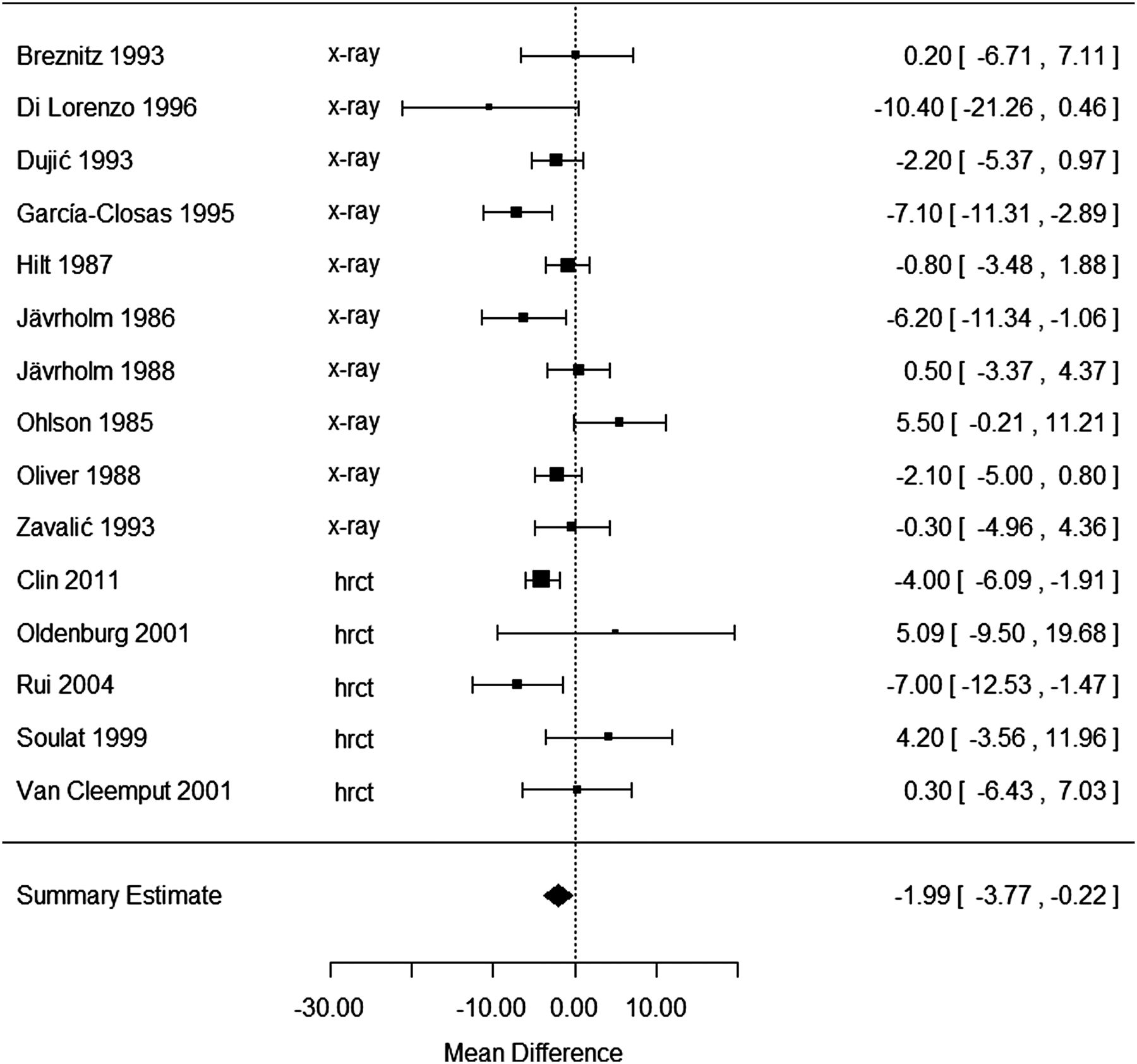
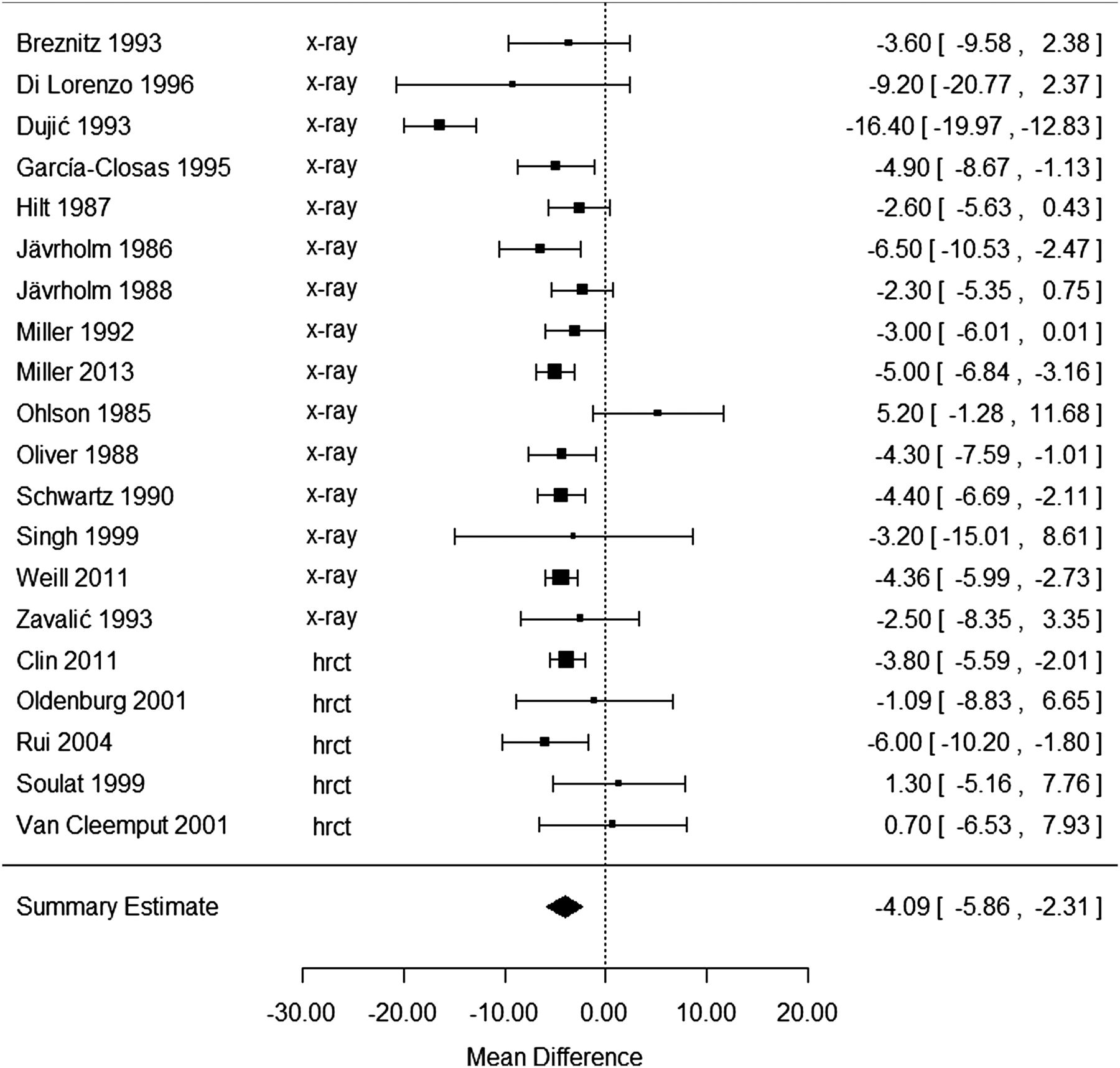
The cross-sectional studies were all conducted among occupationally exposed workers, from a variety of industries (eg, shipbuilding, railroad workers, etc). Study participants were generally male, with mean age at examination of ∼50–60 years. Figure 1 (FVC) figure 2 (FEV1) show individual study results as well as the summary effect estimates resulting from the meta-analyses. The summary effect estimates for FVC and FEV1 are statistically significant, showing a change of −4.09%pred (95% CI −5.86 to −2.31) and −1.99%pred (95% CI −3.77 to −0.22), respectively. The results of larger studies are very consistent in showing a decrease in FVC (see figure 1). In contrast, fewer large studies are available for FEV1, and there is less consistency in the results (see figure 2). The use of random effect models was supported for both pulmonary measures, as the tests for heterogeneity were statistically significant, and the I2 was 80% and 57% for FVC and FEV1, respectively (where I2 represents the proportion of the total variation across studies due to study heterogeneity instead of chance).
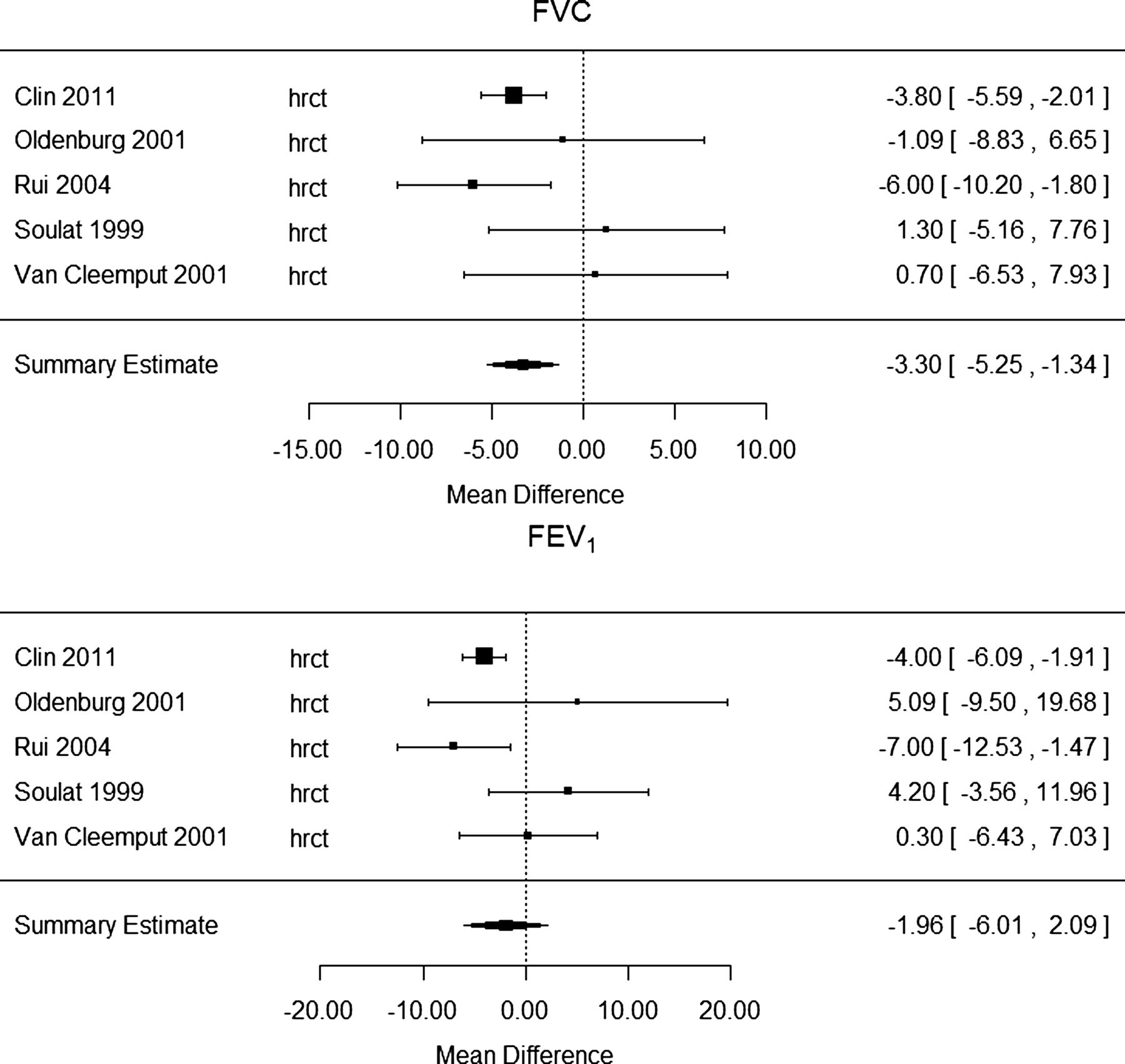
Analysis of the HRCT studies is separately shown in figure 3. Although, the number of study participants varied widely across HRCT studies, for both measures of lung function, results of HRCT studies considered separately are quite similar in magnitude to overall results (combining the two study types) and to X-ray results. For FVC, results from HRCT and X-ray studies considered as separate sets are statistically significant: −3.30%pred (95% CI −5.25 to −1.34) and −4.55%pred (95% CI −6.73 to −2.38), respectively; FEV1 results for HRCT and X-ray studies considered separately were very similar in magnitude to the combined results, but are not statistically significant: −1.96%pred (95% CI −6.01 to 2.09) and −1.87%pred (95% CI −3.96 to 0.23), respectively. Given that the overall (combined) results for FEV1 are statistically significant, this is likely due to the smaller sample sizes when X-ray and HRCT studies are separated. There were no clear asymmetries in the examination of funnel plots (see online supplementary material) for all the analyses (although for HRCT analyses there were few data points) suggesting that publication bias is not an issue in these analyses.

Study-specific and summary effect estimates for change in per cent predicted forced vital capacity comparing asbestos-exposed groups with and without pleural plaques, X-ray and high-resolution CT (HRCT) cross-sectional studies. Data are mean values; bars and values in brackets are 95% CI, size of data point is proportional to study weight.
Study-specific and summary effect estimates for change in per cent predicted FEV1 comparing asbestos-exposed groups with and without pleural plaques, X-ray and high-resolution CT (HRCT) cross-sectional studies. Data are mean values; bars and values in brackets are 95% CI, size of data point is proportional to study weight.

Study-specific and summary effect estimates for change in per cent predicted forced vital capacity (FVC; top panel) and forced expiratory volume in 1 s (FEV1; bottom panel) comparing asbestos-exposed groups with and without pleural plaques, for high-resolution CT (HRCT) cross-sectional studies. Data are mean values; bars and values in brackets are 95% CI, size of data point is proportional to study weight.
For sensitivity analysis, we first excluded studies with the limitations described in the Methods section from the meta-analysis; 16 and 12, respectively, were excluded in the FVC and FEV1 analyses. The results were more consistent (narrower CI despite a smaller number of studies) with a summary effect estimate of −4.08%pred (95% CI −5.44 to −2.71) for FVC (based on four studies25 ,26 ,35 ,38) and an effect for FEV1 that is almost doubled compared with the full set analysis (−3.87%pred, 95% CI −5.84 to −1.90; based on three studies25 ,26 ,38). Next, one study at a time was excluded to evaluate influence of individual studies on the summary effect measures. No one study showed a notable influence on the summary results, which changed by <8% for FVC, and between −18% and +25% for FEV1. In addition, examination of the studies excluded because of analysis or reporting issues (see online supplementary table S2) indicates that the qualitative results of this additional set of studies are also consistent with the pattern seen in figures 1 and 2, with three of the five studies in online supplementary table S2 indicating a decrement in FVC in the pleural plaque group, compared with the no pleural plaque group (two studies did not state if there was a decrease or increase).
Relationship between lung function measures and extent of pleural plaques
Four cross-sectional studies also presented analyses of the extent of pleural plaques in relation to degree of decrement in lung function.22 ,37 ,38 ,48 Lilis et al48 is related to the Miller et al31 study included in the meta-analysis. In the study by Clin et al,38 the decrease in FVC seen with increasing maximum cumulative plaque extent was statistically significant, and for FEV1 the decrease was marginally significant (p=0.06); there was a difference of approximately −4%pred in %pred FVC and %pred FEV1 when comparing the lowest to the highest plaque extent category. In the study by Lilis et al,48 a higher index score (indicating increased pleural plaque size) was significantly associated with a larger decrement of 5–10%pred FVC (accounting for smoking and time since first exposure) compared with a lower index score. Van Cleemput et al22 reported a non-significant decrease in %pred VC and %pred FEV1 with increasing total surface area of pleural plaques; however, on average those with pleural plaques had slightly better lung function than those without pleural plaques. Although van Cleemput et al22 concluded that neither the presence nor the extent of the plaques was correlated with lung function parameters, this is a small study of only 73 workers compared with more than 2000 workers in the study by Clin et al,38 which found that %pred FVC and %pred FEV1 both tended to decrease with increased plaque length. Zavalic et al37 reported that %pred FVC as well as %pred FEV1 tended to become lower with increases in plaque length. Additionally, the longitudinal study by Sichletidis et al49 demonstrated that after 15 years of follow-up, the total surface area of pleural plaques increased twofold and lung function was statistically significantly decreased over that period. Although increased plaque surface area was not statistically significantly associated with the observed reductions in %pred FVC or %pred FEV1, the reduction in total lung capacity was associated with plaque surface area (r=−0.486, p=0.041). Taken together, these studies strongly suggest that the extent of the decrease in lung function is associated with the extent (size or total surface area) of pleural plaques.
Analysis by categorical, rather than continuous measures of lung function
Three studies presented analyses in terms of difference in the proportion of individuals within a group below a specified value for the lung function test or combination of tests. In the study by Oliver et al,34 the proportion with FVC <80%pred was approximately doubled in the pleural plaque group (18.5%) compared with the group with no pleural plaques (9%; relative risk: 2.1, 95% CI 1.1 to 3.7); the smoking-adjusted mean difference between these two groups was −4.3%pred FVC, similar to the summary effect estimate for all studies in our meta-analysis. García-Closas and Christiani28 observed a non-statistically significant increase in the proportion classified as having restrictive disease (defined as FVC <80% predicted and FEV1/FVC >75%), from 3.9% in the group with no pleural plaques to 7.8% in the pleural plaques group. In the study by Dujić et al,27 the estimated relative risk for restrictive disease (defined as FVC <80%pred and FEV1/FVC ≥70%) in the group with pleural plaques, compared with the group with no pleural plaques, was 2.6 (95% CI 1.7 to 3.9); the results in terms of mean difference in %pred FVC between groups in this study were notably larger than other studies in figure 1. The risk of obstructive disease in these studies were not different between those with plaques compared with those without pleural plaques, where obstructive disease was defined as FEV1<80%pred and either FEV1/FVC<70%27 or FEV1/FVC≤75%.28 However, the increase in the proportion of individuals with mixed-pattern disease (FVC and FEV1<80%pred, and 60%<FEV1/FVC<75%), from 1.3% in the no plaques group to 6.5% in the plaques group, was significant in the study by García-Closas and Christiani.28
Evidence that the observed effect is not due to undetected parenchymal changes detectable by HRCT
Analysis of HRCT studies alone showed that undetected parenchymal changes in X-ray examinations (but which would be detectable using HRCT) are not likely to explain the observed effects on lung function. The decrease in FVC observed in HRCT studies was somewhat smaller than that shown in X-ray studies (although still statistically significant); for FEV1 there was little difference in the effect size, although this estimated effect was not statistically significant in the smaller set of HRCT studies.
Analysis of longitudinal studies
Longitudinal studies allow for evaluating the progression of pleural plaques over time, as seen by an increase in the extent of pleural plaques or thickening and a corresponding increase in lung function deficits with the passage of time. Only four longitudinal studies were found in the literature search. The mean length of follow-up varied among these studies from 3.7 to 15 years, with the longer follow-up periods providing evidence supporting an association between pleural plaques and increased rate or degree of pulmonary impairment (see online supplementary table S5). The presence of pleural plaques was not related to differences in decline in %pred FVC or %pred FEV1 measures in the studies with the shortest follow-up (3.7–4 years).24 ,33 In a case–control study with a 7-year follow-up, decreases in FVC of 31±12 (mean±SE) and 15±6 mL/year were seen in those with and without pleural plaques, respectively, but this difference between groups was not statistically significant.50 In the small study of people with plaques only, but with the longest follow-up period, the size of pleural plaques grew more than twofold (from 8.5 to 17.2 cm2) over approximately 15 years,49 and there was a large and statistically significant decrease of 14.6%pred FVC and 4.3%pred FEV1 over the follow-up period. The use of %pred values by Sichletidis et al49 accounts for the expected decline with increasing age over the follow-up period. In addition, the observed pulmonary decrements are unlikely to be the result of continued asbestos exposure; Ostiguy et al50 stated that additional exposure during the follow-up period was low, while Sichletidis et al49 stated that there was no additional exposure during the follow-up period.
Discussion
This systematic review demonstrates statistically significant decrements of 4.09%pred FVC (95% CI 2.31 to 5.86) and 1.99%pred FEV1 (95% CI 0.22 to 3.77) in people exposed to asbestos with pleural plaques relative to exposed people with no pleural plaques. While the total decrement in either group could be due in part to asbestos exposure alone, the estimated difference between the groups should primarily reflect the decrement due to pleural plaques. In the meta-analysis by Wilken et al,8 asbestos-exposed workers without radiological abnormalities showed decrements in lung function (ie, values below 100% of predicted: %pred FVC 95.7 and %pred FEV 93.6). Thus, the lung function decrements associated with pleural plaques in this analysis are even more pronounced when compared with 100%pred, or normal lung function.
Cross-sectional studies also suggest that increased extent of pleural plaques is associated with greater decrements in lung function. Regarding evidence from longitudinal studies; while two of these had very short follow-up periods of less than 5 years,24 ,33 the study with a 15-year follow-up49 showed significant decreases in lung function.
Analysis of HRCT studies alone showed similar results to those of the X-ray studies alone. Thus, undetected parenchymal abnormalities are unlikely to fully account for the lung function decrements observed in X-ray studies. It is also unlikely that the observed association between pleural plaques and lung function decrements reflects solely an independent effect of asbestos exposure on lung function. In this analysis, we compared exposed workers with plaques to exposed workers without plaques: this comparison should reduce the potential influence of differences in exposure on the analysis, although it does not eliminate the possibility that workers with pleural plaques had higher exposure. However, the largest HRCT study controlled for cumulative exposure, as well as other potential confounders, and demonstrated significant pulmonary function decrease consistent with our summary effect estimate.38 Similar results were obtained in a large X-ray study34 that controlled for duration of exposure. A smaller study that stratified for exposure observed a tendency for better lung function among workers with versus without pleural plaques.33 Overall, these results indicate that differences in asbestos exposure are unlikely to fully explain the observed differences in lung function. It is possible, however, that people more sensitive to the effect of asbestos exposure, given the same level of exposure, develop pleural plaques and also have a larger decrease in lung function. In that case, plaques may not be the cause of the decrease in lung function, but are a marker for susceptibility to pulmonary effects of asbestos.
Specific aspects of the design or analysis of these studies indicate that the demonstrated association of pleural plaques and lung function decrease are unlikely to be explained by other causes of lung function loss, such as demographic characteristics, smoking or other lung disease. The sensitivity analysis addressed limitations or potential biases noted through a systematic review of study methods conducted prior to evaluation of the results, including limitations in the way in which smoking was addressed and lack of an explicit statement that some kind of blinding procedure was used for the reading of the X-ray or HRCT. In this sensitivity analysis of studies without limitations in study methods, pulmonary decrements were essentially the same for FVC or increased almost twofold for FEV1 compared with the analysis including all of the studies and the decrements remained statistically significant. Medical reasons for decreases in pulmonary function were explicitly accounted for through exclusion of individuals with lung diseases in seven studies;21 ,23 ,24 ,29 ,30 ,37 ,38 since this type of exclusion is common, it may have been performed but not mentioned in some papers having limited details on participant recruitment and inclusion/exclusion criteria.
The 2000 ILO guidelines define the outcome of LPT as plaques with width of at least 3 mm, a more sensitive and specific definition compared with the 1980 ILO guidelines. Although no studies reported results for plaques with width of at least 3 mm (ie, LPT), one large study38 reported results for plaques less than 2 mm and found that those with such plaques had at least 100%pred FVC and FEV1. Thus, results of this analysis for pleural plaques can be seen to apply to LPT.
We have considered the potential for BMI to affect our observed associations between pleural plaques and lung function. Directionally consistent with this potential bias, we find a tendency for greater FVC decrements in the X-ray studies (4.55%) relative to the HRCT studies (3.30%). However, given that the FVC loss is still observed in the HRCT studies, the associations cannot be fully explained by effects of BMI. Of the two studies that included BMI in their analyses, one study, using X-rays, observed a slightly higher BMI in people with plaques (mean 30.3 and 28.5 kg/m2, respectively for with and without plaques), and higher BMI and age were significantly related to decrements in FVC;36 the results used in our meta-analysis are the BMI-adjusted and age-adjusted results. The other study used HRCT, and observed similar mean BMI between individuals with and without pleural plaques (27.7 and 27.4 kg/m2, respectively).38 More generally, the prediction of FEV1 and FVC is not improved by considering weight after taking into account height, age, race and sex in cross-section analyses of lung function.51 We do not believe large differences in BMI by radiographic group are likely in the remaining studies examined, and overall, we do not believe that our observed associations between pleural plaques and lung function decrements are biased by an effect of BMI.
With regard to fibre type, 13 studies did not report fibre type of asbestos exposure, 4 reported mixed exposure, 2 reported mostly chrysotile exposure and 1 reported Libby tremolite (Libby amphibole asbestos) exposure. Although we could not examine fibre characteristics in this analysis, we are not aware of any studies of pleural plaques and lung function that indicated potential differences in association by fibre. Moreover, the results from the studies included in this meta-analysis did not display great variability, although it is likely that study populations were exposed to different fibre types (or mixtures).
Although the changes in lung function found in our meta-analyses are relatively small (−4.09%pred FVC; −1.99%pred FEV1), these decrements are not inconsequential. The observed decrease in lung function should be considered on an individual as well as a population level. At the individual level, the decrement in FVC or FEV1 may or may not be noticeable for a given patient; while many with pleural plaques could have well-preserved lung function, there are some at the lower end of ‘normal’ lung function, for whom even a small additional decrement would result in an increased disease severity (eg, mild to moderate disease). Thus, at the population level, even small changes in the average of a distribution of lung function can result in a proportion of the exposed population shifted down into the lower ‘tail’ of the lung function distribution. This perspective was noted by the ATS in a 2000 article,52 which stated, “It should be emphasised that a small but significant reduction in a population mean FEV1 or FEV0.75, is probably medically significant, as such a difference may indicate an increase in the number of persons with respiratory impairment in the population. In other words, a small part of the population may manifest a marked change that is medically significant to them, but when diluted with the rest of the population the change appears to be small.” In addition to the mean decrease in the lung function of exposed individuals with pleural plaques, longitudinal studies show a greater rate of decline in the lung function of asbestos exposed individuals with pleural plaques. Both, the mean decrease in lung function we observed and the greater rate of decline in lung function, are consequential from a public health perspective.52
Our review and meta-analyses indicate that pleural plaques (and consequently, LPT as defined in recent ILO guidelines) are associated with declines in lung function. This association is not likely to be fully explained by undetected parenchymal abnormalities. Although the average decrement lung function associated with the presence of pleural plaques may not be generally considered clinically significant, the relation between plaque size and degree of decrement, and the increase in size and decrement over time indicate these changes may be consequential to the exposed population.
Acknowledgments
The authors thank Larissa Pardo for assistance.
References
Supplementary materials
Supplementary Data
This web only file has been produced by the BMJ Publishing Group from an electronic file supplied by the author(s) and has not been edited for content.
Files in this Data Supplement:
- Data supplement 1 - Online supplement
Footnotes
Competing interests None.
Provenance and peer review Not commissioned; externally peer reviewed.